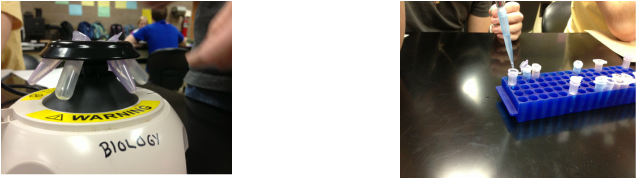
Methods:
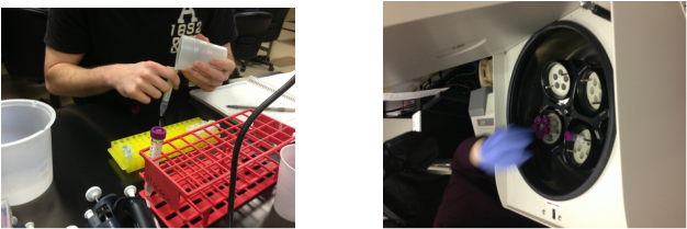
-Methods for DNA extraction: In order to obtain cheek cells, we swished salt solution around in our mouths and expelled the solution into a cup. We then transferred the wash from our cup to a tube provided by instructor, then we capped the tube. The tube was centrifuged in order to create a pellet of matter including DNA at the bottom. When complete, we removed as much supernatant as possible and kept the pellet (containing the DNA). We used a pipette to churn the solution of Chelex several times to suspend the Chelex beads, then quickly transferred the solution into our tube containing the pellet. We thoroughly mixed the components by, again, pipetting the mixture in and out several times. Next, we transferred the mixture to a labeled eppendorf tube and placed a lid lock on the tube. We put the eppendorf tube in a hot block to rupture the cheek cells and release the DNA into solution. Afterwards, we placed the tube on ice to cool and then removed the lid lock so we could separate the DNA from the Chelex and cell debris. Finally, we transferred the extracted DNA supernatant into a new, labeled tube for use in the next step. Any remaining mixture was tossed at the end.
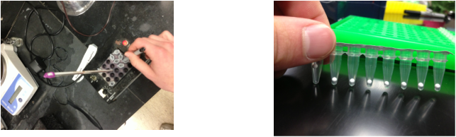
-Methods for PCR: We then conducted the Polymerase Chain Reaction (PCR). First we had to first obtain a PCR tube with a "bead" inside containing the necessary chemicals (Taq DNA polymerase, buffers, and all four deoxyribonucleotides) to carry out the experiment. We placed the primer mix into the tube in order to dissolve the bead. Next we added the extracted DNA to the PCR tube and then mixed the contents. The tube was put into the PCR machine and the DNA was replicated after a few hours.
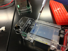
-Methods for Gel Electrophoresis: The agarose gel was precast for each group. In the first well we added a standard 1kb sample ladder of DNA strands for length comparison. Then we added our DNA to each of the next wells, making sure the wells were closest to the negative charge in the tray. The purpose of this was to ensure the negatively charged DNA strands traveled towards the positively charged anode when the electrical current was added. This caused the DNA strands to move through the sponge like gel until some point in the middle of the gel, determined by length of DNA strands and duration of incubation. After adding the current and a 30 minute wait, we had a product we were able to analyze.
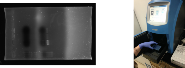
-Methods for Electrophoresis Analysis: We started the analysis by adding a certain dye called Ethidium Bromide to the DNA solution before we ran the electrophoresis. This is a special chemical that lights up the DNA under UV radiation. Once the gel was finished with electrophoresis, we put the gel in a special machine to be able to see the bands even if they are very light. We were able to see just one major band which was our VMAT-2 gene. Once we knew that we could find and see our band of DNA clearly, we had to cut the DNA band out of the agarose gel. We put the gel on a panel of glass over a couple of UV lights to see the band and then used a razor to cut the DNA out. It was important to wear a lot of UV protection from the radiation.
-Methods for DNA Extraction from Gel: The first step taken in this process was cutting out the band of DNA from the agarose gel, which we took care of in our last step. We added QG buffer (based on our gel weight) to the gel in order to dissolve the gel without harming the DNA. Then, the mixture was placed in a 50 degree Celsius hot block to be further dissolved. Afterwards, we introduced isopropanol to our solution to help the DNA stick to the white filter within our column. We transferred our solution to the column to be cleaned by filtering, then centrifuged our samples. The solution at the bottom of the collection tube was discarded, as the DNA was a caught in the white filter of the column. To further clean the samples and remove any excess salt, we washed some QG buffer and PE buffer through our solution. Finally, we added EB to the column in order to wash the DNA through for collection. The DNA (now isolated to the VMAT-2 gene) was then sent to a laboratory at the University of Minnesota to be sequenced.
